
Lab-grown retina reveals secrets of color perception
How cells that allow people to see colors are made? To answer this question, the researchers created a human retina in the laboratory, on which they could freely test their ideas. Research has shown that developing human eyes need an appropriate hormone in order to see colors properly. This research could help develop treatments for diseases such as color blindness and macular degeneration.
In laboratories belonging to the Johns Hopkins University in Baltimore, scientists have grown human retinas from scratch ka. They wanted to make this way b to learn the secret of the formation of com color-perceiving records. Their research has been published in the journal „Science”.
Scientists conducted research on the so-called. organoids. These are miniature versions of r organs In this case, the net retinas, grown from com stem cells, which are responsible for the perception of blue The cells retain their key anatomical features. These small, tr 3D models are an invaluable aid for research. Most eye research is done on mice and fish, but none of these species The retina is not as colorful as a human. Trichromatic color vision in the odr The number of trees in Poland is at the level in, so m.in. the researchers were working on organoids.
– Everything we study looks like a normal human eye, only developing in a dish – said Robert Johnston of Johns Hopkins University. – Such a model can be manipulated without directly interfering with the human body – added.
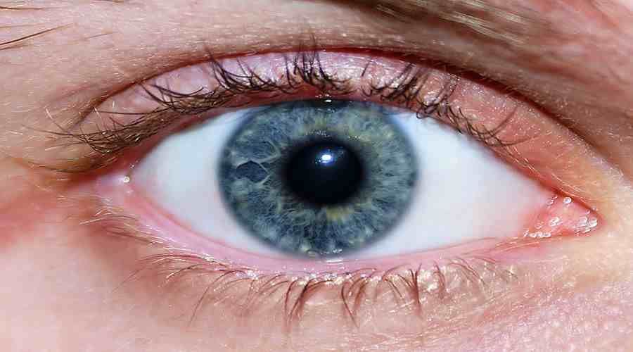
The researchers were interested in how b determines the fate of the cell rki. What transforms the developing com tion in a specific type of light-absorbing cells, i.e rki. This aspect of human biology is largely unknown. Scientists have focused on specialised com The light-absorbing cells, i.e., the photoreceptors, or more precisely, the three photoreceptors – the so-called “light-sensitive receptors”. cones, which that allow us to see the colors blue, red and green.
During the development of the retinal organoid ki, scientists have observed that the first to appear were cells The blue-perceiving cells in the brain. Then came the com rks that detect red and green colors. It turned out that the key to the molecular change was a thyroid hormone. Importantly, the levels of this hormone were not controlled by the thyroid gland, which of course, was not in the dish, but through the eye itself.
The amount of thyroid hormone dictated how a cell would behave rki. By understanding this process, the researchers were able to manipulate the development of the reticular in the cells, creating those who If they were part of the human eye, they would only be able to perceive the color blue, or only green or red.
The discovery may explain why in premature babies in, which those with low levels of thyroid hormone were more likely to have visual disturbances. – If we can answer the question of what is driving the com to its final form, we are closer to being able to restore of color vision to people who ho have damaged photoreceptors – indicated Kiara Eldred, the head The author of the study.
But this is just the beginning of scientists’ research from the Johns Hopkins University. In the future, they want to use the organoid, of course to learn even more about color vision and the mechanisms involved in creating other area in the grid of the speck, such as the of the retina. Macular degeneration ltej is one of the main of the various causes of blindness in humans. Understanding how to create a new speck of ¿ The results could lead to new and effective therapies.
– Our work establishes human organoids as a model system for studying the mechanism in human evolution. This is exciting. Organoids take nine months to develop, just like a human baby. So what we’re really investigating is the development a fetus – Johnston admitted.